Better Outcomes With AI-Based Medical Imaging

The vast majority of today’s healthcare data comes from medical scans, and doctors have become stressed and overburdened as they struggle to interpret the images while managing patient care. By using AI and deep-learning technology to analyze patient scans, doctors can obtain results much faster while also improving diagnostic accuracy.
Scans are not as easy to decipher as they may appear. Many contain dozens of images that doctors must pore over to arrive at a diagnosis. Pinpointing the exact location and dimensions of fractures, nodules, and other lesions is often difficult.
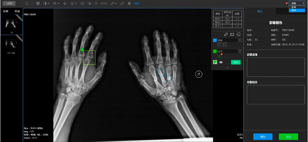
When AI algorithms analyze scans, they can quickly point doctors to the location of a fracture or nodule, saving time (Figure 1). “We call it AI-assisted diagnosis,” says Xiangfei Chai, founder and CEO of HY Medical, a Beijing-based company that develops AI-based imaging solutions. “Doctors still make the decision, but they can do it two to three times faster than they can with traditional scanning.”

AI-based scanning analyzes patient image data to uncover characteristics that can’t be seen by the human eye. Both these features help doctors improve the accuracy of their diagnoses. “Our AI-Assisted Diagnosis of Medical Imaging Solution can increase accuracy up to 15%,” Chai says.
“Our #AI-Assisted Diagnosis of #Medical Imaging Solution can increase diagnosis accuracy up to 15%.” —Xiangfei Chai, CEO of HY Medical via @insightdottech
AI Medical Imaging for Surgical Decisions
The HY solution has been working with more than 10 diseases so far, such as fractures, aortic dissection, aortic abdominal aneurism, and some cancers. The experience of one of Beijing’s largest hospitals shows how its precise calculations can make a difference.
To treat aortic dissection—a tear in the inner layer of the large blood vessel coming from the heart—doctors often use a stent. Stents come in several sizes, and the blood vessel’s “U” shape makes measurement of the tear’s dimensions difficult.
At the Beijing hospital, an HY Medical study found that almost half of stent surgeries failed the first time, and nearly 20% of the time, surgeons used a wrong-size stent. After the hospital adopted HY’s AI-based solution, stent selection accuracy increased by 50%. The solution also provided results within 10 minutes, decreasing waiting time for doctors and patients.
AI-Enabled Medical Imaging as a Disease Management Tool
AI can also help doctors segregate patients according to disease severity. That capability was helpful when the COVID-19 pandemic broke out in China.
Initially, there was a shortage of tests for the virus. In addition, early tests generated many false positives—at a time when providers were overwhelmed. As a result, some doctors switched to doing CT scans instead. Machines enhanced with the HY solution’s AI capabilities not only accurately diagnosed the disease, but revealed how sick people were, allowing hospitals to effectively triage the deluge of patients.
Later, as lab tests became more accessible and reliable, doctors used AI scanning to track the progress of patients’ infections.
“AI automatically calculates the size of a lesion and how fast it is shrinking or growing,” Chai says. “It also predicts the course of development based on the lesion’s rate of change, allowing doctors to provide personalized treatment.”
When the pandemic spread beyond China, it hit the U.K. particularly hard. HY worked closely with scientists at Coventry University to quickly develop an AI-enabled scanning solution tailored to the specific needs of U.K. doctors. They put the solution to work in the university’s affiliated hospital, where it was downloaded within 24 hours, giving physicians the ability to tend to the most severe cases right away.
“There are so many uncertainties during the treatment of a COVID-19 patient, such as what treatment should be applied or when the patient should be sent to the ICU,” Chai says. “With AI technology, better decisions can be made about assessing which patients really needs beds and these who can safely go home.”
Expanding to Treatment
As information about AI-based imaging’s efficiency and accuracy spreads, more providers are incorporating the technology.
To give clinicians flexibility, HY provides three ways to deploy its solution: Doctors can upload scanned images to a cloud-based app, large hospitals can connect the solution to their internal imaging networks, and smaller hospitals with a single X-ray machine or other scanning device can load the HY appliance directly onto the machine. All calculations are done by Intel® Xeon® Scalable processor-based systems.
As more hospitals incorporate AI-enabled imaging, it is expanding to new use cases in surgery, radiation oncology, and chemotherapy.
To help providers build these applications, HY’s solution uses the Intel® OpenVINO™ Toolkit, which allows developers to easily transport code from one application to another and tweak it to accommodate particular needs. “You just do a little bit of setup—you don’t need to change the algorithm much to adapt,” Chai says.
The more hospitals use AI algorithms, the more that data iterates, improving accuracy. And as AI evolves, it will increase in use to predict treatment outcomes.
For example, HY is working on an imaging solution that will judge whether individual breast cancer patients are likely to respond well or poorly to various forms of chemotherapy. It is also developing predictive algorithms for cancer and artery disease.
“Worldwide, there are many groups working on predictive solutions and other new applications,” Chai says. “In the future, AI will have a much bigger influence on medicine.”